
 80813-00200


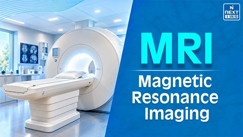
The full form of MRI is Magnetic Resonance Imaging (मैग्नेटिक रेजोनेंस इमेजिंग). It uses strong magnetic fields and radio waves to generate high resolution 3D images of soft tissues without using harmful ionising radiation. Interestingly, the MRI scanners use liquid helium to supercool the superconducting electromagnets and generate magnetic fields about 30,000 times stronger than Earth's gravity. The MRI machine aligns hydrogen atoms in the human body. The radio pulses emit signals as they re-align to map organs, brain and ligaments.
How MRI Works?
- Magnetic Resonance Imaging or MRI uses a powerful magnetic field, radio waves and a computer to create a detailed image of the body. The MRI works primarily by detecting signals from hydrogen nuclei because the human body contains a large amount of water and fat.
- The radio frequency pulses temporarily disturb this alignment. When the pulses stop, the protons return to their original state and release the signals. The receiver coils detect these signals and the magnetic field tracks the location of signals.
- Various tissues release signals at different frequencies. Thus the computer converts them into contrasting images. The MRI may use special sequence or gadolinium contrast to highlight particular tissues, blood vessels or abnormalities. It does not use ionising radiation.
Applications & Uses of Magnetic Resonance Imaging
| S.No. | Field | Use of MRI |
|---|---|---|
| 1 | Materials science |
|
| 2 | Chemical research |
|
| 3 | Food science |
|
| 4 | Agriculture |
|
| 5 | Archaeology |
|
| 6 | Petroleum industry |
|
| 7 | Engineering |
|
| 8 | Pharmaceutical research |
|
| 9 | Forensic science |
|
| 10 | Veterinary science |
|
What is an MRI Test?
An MRI test is a painless, non-invasive imaging scan or test which uses powerful magnetic fields, radio waves and computer processing to produce detailed pictures of organs, tissues and other body parts. During an MRI scan or test, the patient lays down on a movable table that slides onto a tunnel-shaped scanner (in case of Closed MRI system). The patients are asked not to move their body during the image capture by MRI machine. Sometimes, a contrast agent is injected into the body to improve the image quality and clarity. An MRI generally takes about 30 to 60 minutes and doesn't make use of X-ray radiation.
Comparison of Open and Closed MRI
| S.No. | Feature | Open MRI | Closed MRI |
|---|---|---|---|
| 1 | Design |
|
|
| 2 | Patient comfort |
|
|
| 3 | Magnetic strength |
|
|
| 4 | Image quality |
|
|
| 5 | Scan duration |
|
|
| 6 | Suitable for |
|
|
| 7 | Availability |
|
|
| 8 | Limitations |
|
|
History
Magnetic Resonance Imaging (MRI) emerged from nuclear magnetic resonance research which was initially used for chemical analysis. In the early 1970s, Paul C. Lauterbur had introduced magnetic-field gradients that enabled signals from hydrogen nuclei to be converted into images. Afterwards, Peter Mansfield has developed faster ways for acquiring signals and creating clear images. Various early experiments produced images of body parts such as the hand and finger before the technology became advanced for clinical use.
Over the period of time, MRI has developed into an important diagnostic tool because it can provide detailed images of soft tissues without using ionizing radiation. In 2003, Lauterbur and Mansfield received the Nobel Prize in Physiology or Medicine for discoveries related to MRI.
Difference Between MRI and CT Scan
| S.No. | Basis | MRI | CT scan |
|---|---|---|---|
| 1 | Full form |
|
|
| 2 | Main technology |
|
|
| 3 | Ionising radiation |
|
|
| 4 | Soft-tissue detail |
|
|
| 5 | Bone imaging |
|
|
| 6 | Speed |
|
|
| 7 | Noise |
|
|
| 8 | Claustrophobia |
|
|
| 9 | Metal and implants |
|
|
| 10 | Contrast agent |
|
|
| 11 | Common uses |
|
|
FAQs about MRI
What is the full form of MRI?
The MRI full form is Magnetic Resonance Imaging. It is a technique to take images and uses magnet and radio waves to study the soft tissues inside the body.
What is the cost of an MRI?
The cost of MRI varies by body parts, facility, and location. The patients should confirm the charges with the diagnostic centres.
What is an MRI test?
An MRI test is an imaging test or scan which creates a detailed image of organs, tissues and body structures.
What is an MRI scan used to diagnose?
An MRI scan is used to diagnose brain, spine, soft tissue, vascular, abdominal, pelvic and tumour-related conditions.
Is MRI safe for patients?
Yes, an MRI is usually safe because it does not use X-rays but the implant and metal screening is essential.
